Introduction
DAPI, or 4’,6-diamidino-2-phenylindole, is a fluorescent stain commonly used in fluorescence microscopy to label DNA. Understanding the principles of DAPI excitation and emission is crucial for researchers aiming to visualize and analyze cellular structures. This article delves into the mechanics of DAPI excitation emission, highlighting their significance and applications in biological research.
What is DAPI?
DAPI is a blue fluorescent dye that binds selectively to the adenine-thymine rich regions in DNA. It is widely used to stain the nuclei of cells, providing a clear view of nuclear structures. Upon binding to DNA, DAPI exhibits strong fluorescence, which can be detected using specific excitation and emission filters in a fluorescence microscope.
Principles of DAPI Excitation and Emission
Fluorescence involves the absorption of light at a specific wavelength (excitation) and the subsequent emission of light at a different, typically longer wavelength. For DAPI:
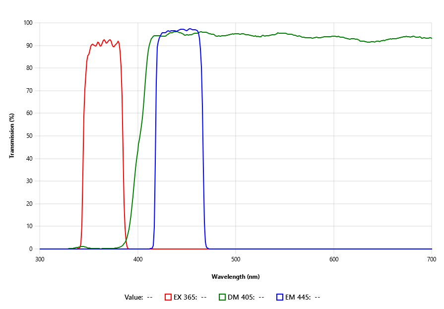
Excitation: DAPI is excited by ultraviolet light, with a maximum excitation wavelength around 358 nm. When DAPI molecules absorb this light, they are raised to a higher energy state.
Emission: As the excited DAPI molecules return to their ground state, they emit light at a longer wavelength, around 461 nm, which appears blue. This emitted light is then captured to produce fluorescence images.
The Role of Excitation and Emission Filters
In fluorescence microscopy, the use of specific filters is essential to isolate the excitation and emission wavelengths of DAPI:
Excitation Filter: This filter allows only the ultraviolet light needed to excite DAPI to pass through, ensuring that the dye is efficiently excited without interference from other wavelengths.
Emission Filter: This filter blocks all light except for the blue fluorescence emitted by DAPI, providing a clear and specific image of the stained nuclei.
Applications of DAPI in Research
DAPI staining, combined with appropriate excitation and emission filters, is a powerful tool in various research fields:
Cell Biology: DAPI is used to visualize cell nuclei, aiding in the study of cell cycle dynamics, apoptosis, and nuclear organization.
Histology: DAPI staining helps in identifying nuclear morphology in tissue sections, facilitating the analysis of tissue structure and pathology.
Genetics: Researchers use DAPI to stain chromosomes in karyotyping, allowing for the identification of chromosomal abnormalities.
Medical Diagnostics: In diagnostic laboratories, DAPI staining is employed to detect abnormal cells, such as cancerous cells, in tissue samples.
Advantages of DAPI Staining
The use of DAPI and its excitation and emission filters offers several benefits:
High Specificity: The filters ensure that only the desired wavelengths are used, reducing background noise and enhancing image clarity.
Enhanced Contrast: By isolating the blue fluorescence of DAPI, these filters provide high contrast images, making it easier to distinguish stained nuclei from the surrounding structures.
Reliable Results: Accurate filtering leads to reproducible and reliable results, essential for scientific research and diagnostics.
Conclusion
Understanding DAPI excitation and emission is fundamental for effectively using this fluorescent dye in microscopy. The specific excitation and emission filters play a crucial role in obtaining clear and precise images of DAPI-stained nuclei. Whether in cell biology, histology, genetics, or medical diagnostics, DAPI remains an invaluable tool for researchers, providing critical insights into cellular and molecular structures.