The C-arm X-ray machine, exemplified by Siemens' model, features a C-shaped design connecting X-ray sources and detectors. Widely used in orthopedic, urology, and cardiac surgeries, it offers real-time imaging and surgical navigation, aided by its X-ray generator, image intensifier, and workstation.
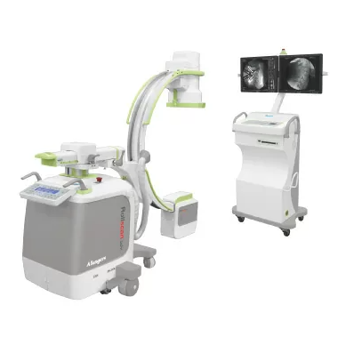
The C Arm X-ray machine is an advanced medical imaging device. It is primarily based on X-ray technology however backed with more efficient and advancing imaging. They are used for fluoroscopy capability – well they also have radiography capability too.
Siemens's C-arm machineis called the ‘C arm’ for its C-shaped arms. It is used to connect the X-ray sources on one end and the detector on the other end.
Usage of C-Arm X-Ray Machine
These machines are widely used for orthopedic, urology, and gastroenterology surgeries. In fact, nowadays, doctors are using it for complicated surgeries including pain management and emergency procedures like cardiac and angiography studies.
With the help of Fluoroscopy technology doctors are able to get high-resolution X-ray images – so that they can do accurate procedures with better outcomes. The systems provide a great deal of flexibility to the surgeon and can take images of the full body.
· The application of a C-arm X-ray machine is not limited to the digestive, cardiac, reproductive, and blood circulation system.
· Doctors can view real-time gallbladder, liver, bone, and several structures. In addition, multiple views are possible.
· Surgical navigation is one of the crucial applications to aid in verifying the placement of all types of implant devices.
· It can also guide needle placement procedures especially when injecting anesthetic medicines.
Key Components
It comprises of X-ray generator with an image intensifier (flat panel detector) and a workstation. It comes with a control and setting option. The C-shaped arm connects the generator and allows the movement horizontally around the swivel axes. These procedures help doctors to take X-ray images from many angles.
The generator emits an X-ray penetrating the body which is placed in between. The intensifier converts the X-ray into a visible image displayed on the C-arm monitor.
Let’s delve into the mechanism:
X-Ray Generator
It is typically placed inside the frame when the C-arm is mounted. Well, it is controlled by the workstation unit. Doctors can modify the operations of the system in real time. With the increasing X-ray power, there is complete flexibility. Since the exposure time is less there is hardly any risk involved.
Image System
The c-arm X-ray machine is backed with a robust imaging system that has the ability to perform multiple movements in single procedures. This advantage comes in handy during various surgical procedures including orthopedic, urology, and cardiology. The entire system is very compact and light in weight. Hence, it allows multiple positioning along with a wide range of motion. The best part is there is hardly any possibility of misalignment between the procedures.
Workstation Unit
The entire operation of the C-arm system is managed with the workstation unit. It contains several handles enabling movements and positioning, and switches – it controls the power supply, light exposure, cable hanger, and control for radiographic setting.